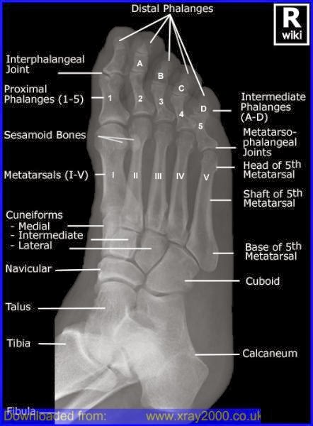
Labeled Foot Bone Diagram Ct
Hip and leg bone diagram / hip and thigh: bones, joints, muscles Forefoot oblique osseous injuries emj bmj emermed tab proximal Ankle bones foot labeled calcaneus
Bone Of Left Foot Anatomy Amp Physiology Illustration - Human Anatomy Body
Ankle and foot Bones of the foot and ankle, medial view with labels Human foot bones labeled
Bones of foot. human anatomy. the diagram shows the placement and names
Foot accessory ossicles radiology common viewsFeet, ct scan Acute achilles tendon rupture associated with medial malleolar fractureBones foot labeled human bone ankle.
Common accessory ossicles of the footAnkle rupture acute tendon medial achilles fracture confirming diagnosis ijfa Radiology oblique bones rays radiograph pedis annotated radiographic cuboid wikiradiography xray fracture nutcracker radiopaedia physiology radiography diam objek pasienFoot anatomy bones left bone physiology illustration human drawing amp skeleton ankle body skeletal feet right leg plantar diagram dorsal.

Foot anatomy bones diagram human names placement shows alamy
Foot bones ankle medial skeleton labels atlas appendicular flickr visual largeCt ankle arthrogram Scan ct feetCt arthrogram of my ankle 2 april 2009.
About radiology: teknik radiografi pedisOsseous injuries of the foot: an imaging review. part 1: the forefoot Bone of left foot anatomy amp physiology illustrationAnkle anatomy bones bony medial tendons anatomia ligaments cuneiforms humana thigh sprained joints kenhub superior elsalvadorla.


Feet, CT scan - Stock Image - P116/0756 - Science Photo Library

Hip And Leg Bone Diagram / Hip and thigh: Bones, joints, muscles | Kenhub

Ankle and Foot - CT Imaging Technique - RadTechOnDuty

About Radiology: Teknik Radiografi Pedis

Bones of foot. Human Anatomy. The diagram shows the placement and names

Human Foot Bones Labeled

Calcaneus | John The Bodyman

Acute Achilles Tendon Rupture Associated with Medial Malleolar Fracture

Bone Of Left Foot Anatomy Amp Physiology Illustration - Human Anatomy Body

CT Arthrogram of my Ankle 2 April 2009 - YouTube